
Advanced Dental Imaging & Digital X-Ray
3D Digital Cone-Beam X-Ray (CBCT)
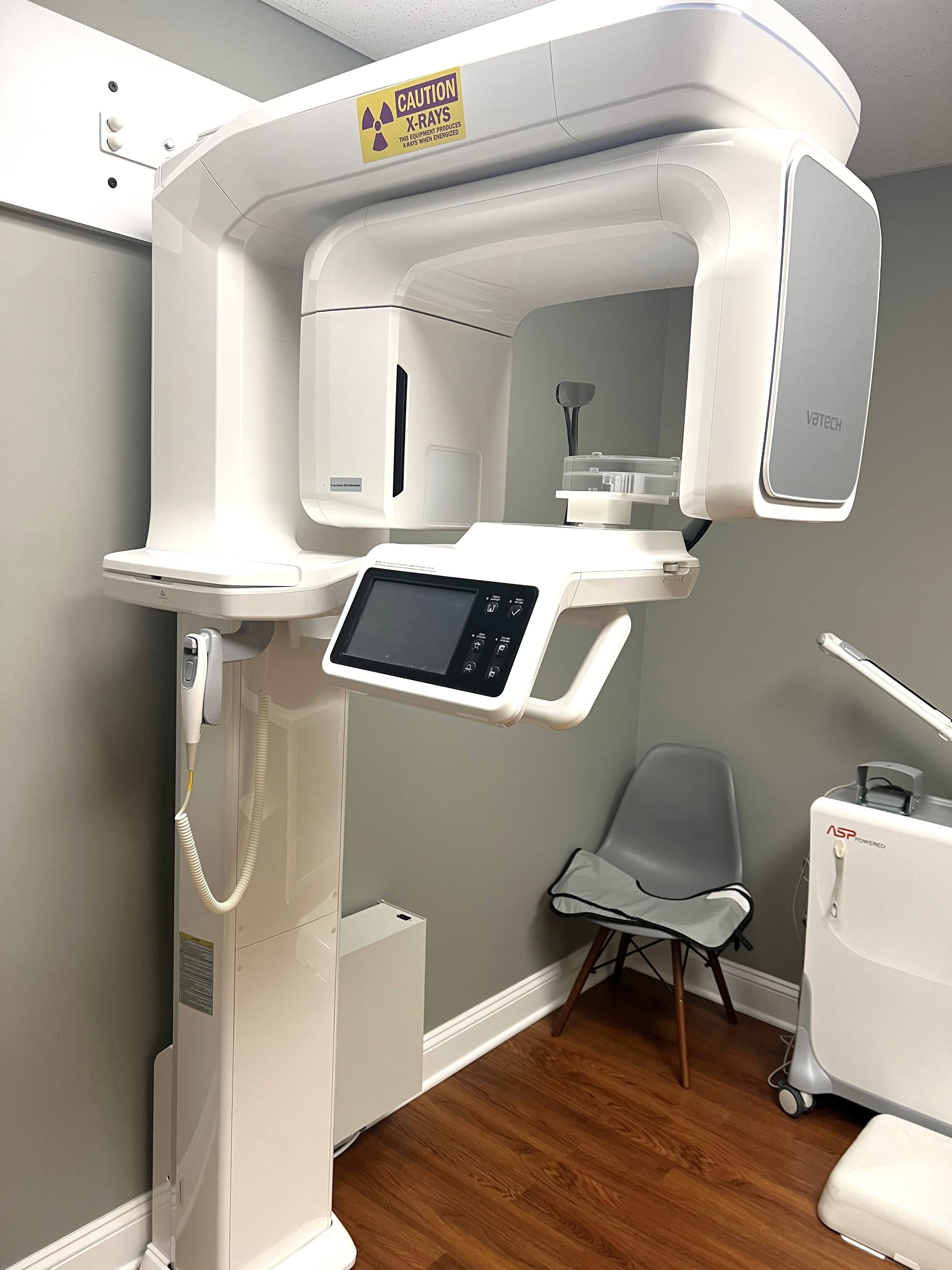
Our cone-beam system captures high-resolution 3D images of the teeth, bone, and surrounding anatomy. This tool plays a key role in evaluating complex cases, planning dental implants, and assessing bone loss from periodontal disease.
Benefits:
Detailed Imaging: Provides a complete three-dimensional view of tooth and bone structures that cannot be seen with traditional 2D x-rays.
Treatment Precision: Helps guide implant placement, assess bone density, and avoid important anatomical structures like nerves and sinuses.
Efficient & Safe: Scans are fast, comfortable, and use significantly less radiation than medical CT scans.
Wall-Mounted Intraoral Digital X-ray Machine

Our practice uses a modern wall-mounted intraoral x-ray system to capture highly detailed images of your teeth and supporting bone as you recline in comfort. The x-ray head is easily positioned at any angle and distance, allowing us to take precise images quickly and safely.
Benefits:
Safety: Although traditional dental films require very little radiation for image development, our digital x-ray system provides unsurpassed safety: a ~90% reduction compared to most older film-based systems.
Time Savings: Images captured instantly with a digital sensor lessen the time you spend in the dental chair.
Better Outcomes: Improved image clarity increases Dr. Kelner's ability to diagnose and treat conditions. Software tools bring detailed magnification and detection tools into focus.
Intra-Oral Camera

Intra-oral imaging gives you a detailed view of your mouth in pictures and videos. Putting specific problem areas onto a computer screen lets us see with a clarity and detail not possible with a traditional mouth mirror. Specialized handheld wands capture the details of your teeth and gums. Even small cracks that often lead to broken teeth appear vividly, with the click of a button.